What Is Hydrocephalus? Symptoms Treatment Options What to Expect Request Consultation
Interactive Patient Education

Understanding
Hydrocephalus
and How It Is Treated

Hydrocephalus is a buildup of the fluid that normally cushions the brain. When that fluid cannot drain and absorb the way it should, pressure rises inside the head. It affects newborns, children, and adults, and it is very treatable. This page walks you through what is happening, how we treat it, and what to expect.

Coronal view of the brain in hydrocephalus, showing the enlarged fluid-filled ventricles highlighted at the center
In hydrocephalus, trapped fluid enlarges the ventricles (shown in red) and raises pressure inside the head.
What Is Hydrocephalus?

A problem of fluid balance in the brain — not simply "water on the brain."

Deep inside the brain are four connected, fluid-filled spaces called ventricles. They produce and hold a clear liquid called cerebrospinal fluid (CSF). This fluid cushions the brain and spinal cord, delivers nutrients, and carries away waste. In a healthy brain, CSF is made continuously, flows through the ventricles and around the brain and spinal cord, and is absorbed back into the bloodstream at about the same rate it is produced.

Hydrocephalus develops when that balance is disrupted — because a passage is blocked, because too much fluid is made, or because the fluid is not absorbed well enough. CSF then backs up, the ventricles enlarge, and pressure builds inside the skull. Over time, that pressure can injure the brain, which is why hydrocephalus is treated rather than simply watched.

The goal of treatment is straightforward: give the trapped fluid a reliable way out so the pressure returns to normal. At Brown Neurosurgery, that may mean a shunt that drains fluid to another part of the body, or, for the right patient, an endoscopic procedure that creates a new internal drainage route without any implanted hardware. Which approach fits best depends on the cause, the patient's age, and the anatomy.

Key Facts
What builds up: Cerebrospinal fluid (CSF)
Where: The brain's ventricles
Who it affects: All ages — infants to older adults
Main treatments: Shunt or ETV
Is it curable? Controllable, not cured — lifelong follow-up
Emergency signs: See "Symptoms" below
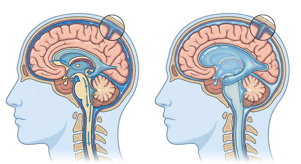
Sagittal (side) view of the head comparing normal cerebrospinal fluid circulation on the left with enlarged ventricles from hydrocephalus on the right
Left: normal CSF flow — fluid is made in the ventricles, circulates through the brain and around the spinal cord, and is absorbed back into the bloodstream. Right: in hydrocephalus, that flow or absorption is disrupted, fluid backs up, and the ventricles enlarge.

Not All Hydrocephalus Is the Same

The type matters because it guides which treatment is likely to work. These are the categories your team will use.

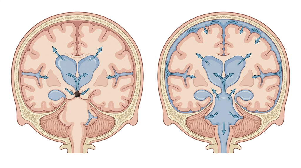
Two front-view brain cross-sections comparing obstructive hydrocephalus, with a blockage, to communicating hydrocephalus, with open pathways but poor reabsorption
Left: an obstructive (noncommunicating) blockage traps fluid and enlarges the ventricles above it. Right: in communicating hydrocephalus the pathways stay open, but fluid is not reabsorbed well, so the ventricles still enlarge.

Communicating

The passages between the ventricles stay open, but the fluid is not absorbed back into the bloodstream fast enough (or, less often, too much is made). Fluid backs up even though it can still flow freely.

Noncommunicating (obstructive)

A blockage inside the narrow channels of the brain stops CSF from leaving the ventricles, so it collects behind the blockage. A common example is a blockage of the narrow aqueduct connecting the third and fourth ventricles.

Congenital & acquired

Congenital hydrocephalus is present at or before birth. Acquired hydrocephalus develops later — after bleeding in the brain, an infection such as meningitis, a head injury, or a tumor. In most cases the cause is beyond a person's control.

Normal pressure hydrocephalus (NPH)

Seen mainly in older adults, NPH enlarges the ventricles with little or no rise in measured pressure. It causes a classic triad: changes in walking, mild memory and thinking problems, and loss of bladder control. It matters because these symptoms can improve with treatment and are sometimes mistaken for other conditions.

What Hydrocephalus Looks Like — by Age

Symptoms depend a great deal on age, because a baby's skull can still expand while an adult's cannot. Choose an age group, then select a sign to learn more.

Select a sign at left to see what it means and when it needs urgent attention.

When to seek care urgently: a rapidly worsening headache, repeated vomiting, increasing sleepiness or confusion, new vision problems, or — in a baby — a bulging soft spot and a fast-growing head. If you or your child already has a shunt and these appear, contact your neurosurgery team right away or seek emergency care, because they can signal a shunt problem.

How Hydrocephalus Is Diagnosed

The evaluation confirms the diagnosis, shows the size of the ventricles, and — for some patients — helps predict whether draining fluid will help.

Imaging

In babies with an open soft spot, a painless ultrasound can image the ventricles. A CT scan quickly shows their size, and MRI gives detailed pictures that can reveal a cause such as a blockage or tumor and show how fluid is moving.

Examination & history

Your team reviews symptoms and their timeline, checks a baby's head measurements against normal growth curves, and performs a neurological exam. In children, faltering development is an important clue.

Predicting shunt response (NPH)

For suspected NPH, removing a modest amount of spinal fluid with a lumbar puncture — or draining fluid for a few days — and watching whether walking improves helps predict who is likely to benefit from a shunt.

Planning the approach

The imaging also shows the anatomy the surgeon needs to choose between a shunt and an endoscopic procedure — for example, whether a single blockage is causing the problem and whether the ventricles are shaped favorably for ETV.

Two Ways to Restore Normal Drainage

Both approaches relieve the pressure by giving CSF a way out. They suit different situations, and your surgeon will explain which fits you.

Shunt

A permanent internal drainage system
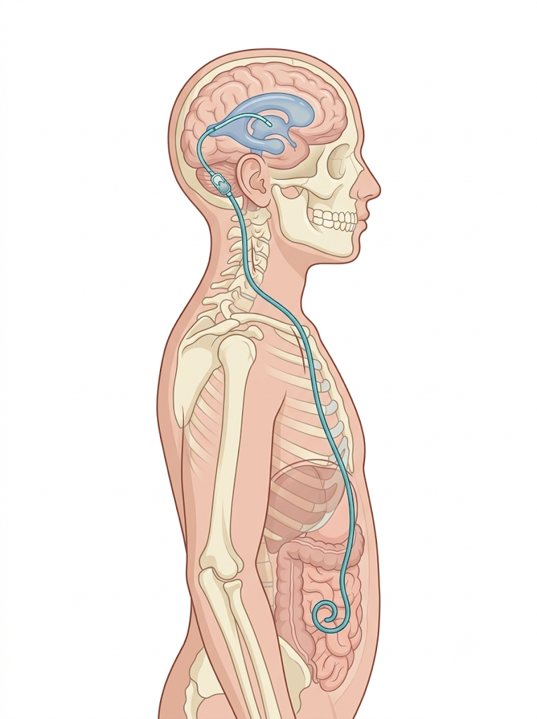
A ventriculoperitoneal shunt draining fluid from a brain ventricle through tubing under the skin to the abdomen
What it isA soft tube with a one-way valve, placed entirely under the skin, that drains CSF from a ventricle to another part of the body — most often the abdomen (a ventriculoperitoneal, or VP, shunt), sometimes the heart.
Best forMost types of hydrocephalus, including communicating hydrocephalus and NPH, and patients not suited to ETV.
Trade-offHighly effective and adjustable, but it is implanted hardware that can, over time, block or become infected and may need revision.

ETV

Endoscopic third ventriculostomy
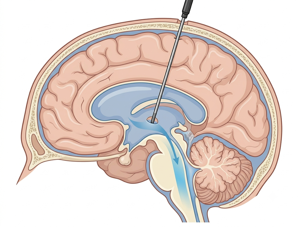
An endoscope creating an opening in the floor of the third ventricle so fluid can bypass a blockage
What it isUsing a tiny camera passed into the ventricle, the surgeon makes a small opening in the floor of the third ventricle so trapped fluid can bypass the blockage and drain the way it normally would. No hardware is left behind.
Best forSelected patients with a blockage-type (obstructive) hydrocephalus and favorable anatomy; success also depends on age and cause.
Trade-offAvoids a lifelong implant, but the new opening can close over time, and it is not suitable for every type of hydrocephalus.
A shunt controls hydrocephalus; it does not cure it. Either way, hydrocephalus is usually a lifelong condition, so ongoing follow-up with your neurosurgery team is part of the plan — to keep the drainage working and to catch any problem early.

Shunt Surgery, Step by Step

Select each step to see what happens and why. Shunt placement is a relatively short operation done under general anesthesia by a neurosurgeon.

Shunt Valves — and an Important Note About MRI

The valve is the part of a shunt that controls how much fluid drains. There are a few kinds, and one has a safety point worth knowing.

Fixed-pressure valve

Opens at one preset pressure chosen by the surgeon. Simple and reliable, but the setting cannot be changed later — a different setting would require a small operation to exchange the valve.

Flow-regulating valve

Aims to keep the amount of fluid draining roughly steady across a range of pressures, closer to the body's natural rate, and can drain faster if pressure rises sharply.

Programmable (adjustable) valve

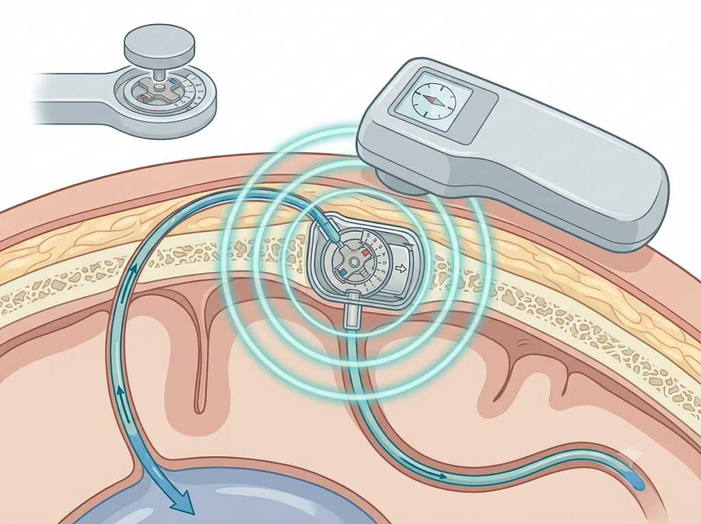
Lets the surgeon change the opening pressure without surgery, using a small external device held against the skin over the valve. The adjustment takes seconds in the office and can be repeated to fine-tune your treatment.

Why the type matters day to day

A programmable valve means many drainage problems can be corrected in clinic, with no operation — one of the reasons they are widely used. But it also means the setting must be protected from strong magnets.

A programmable shunt valve implanted under the scalp being adjusted non-invasively by a handheld programmer held above the skin
A programmable valve’s opening pressure is changed without surgery: a handheld programmer held against the scalp resets the valve beneath the skin using a magnetic signal. The inset shows the valve’s adjustable dial.
Programmable valves and MRI: the strong magnetic field of an MRI scanner can change the setting of some programmable valves. Newer valves are built to resist this, but as a safety step your care team should check and confirm the valve setting after any MRI. Always tell imaging staff you have a programmable shunt, and keep any device card your team gives you.

What Outcomes Can Patients Expect?

Treatment reliably relieves pressure and, for many, restores lost function. Being honest about the trade-offs helps you plan.

Fast relief
Draining the trapped fluid lowers the pressure quickly; pressure symptoms such as headache and drowsiness often ease within days.
Lifelong
Hydrocephalus is a chronic condition. A shunt manages it; regular follow-up keeps it working over years.
Revisions happen
Shunts can block, disconnect, or become infected and sometimes need revision — most common in the first years and in young children.

Risks and Complications to Understand

The main shunt complications are obstruction (a blockage that lets the original symptoms return), infection (redness, swelling or tenderness along the shunt track, often with fever — most likely soon after surgery), and overdrainage (removing too much fluid, which can cause headaches or, rarely, bleeding around the brain). Tubing can also disconnect or a valve can malfunction. With ETV, the new opening can close over time and symptoms can recur, sometimes leading to a shunt later. For NPH, improvement in walking, memory, and bladder control varies from person to person and is more likely when careful testing suggested a good chance of response. Your surgeon will review your individual risks and expected benefit in detail.

Frequently Asked Questions

Does a shunt cure hydrocephalus?
A shunt controls hydrocephalus and relieves the pressure, but it does not cure the underlying condition. Hydrocephalus is usually lifelong, so ongoing follow-up with your neurosurgery team matters. With a well-functioning shunt, most people lead full, active lives.
Will the shunt be visible or limit activities?
The entire shunt sits under the skin, with no external parts. Most people are not restricted in daily activities, though your surgeon may advise against certain high-impact or extreme activities. Your team will give guidance based on your situation.
How is a programmable valve adjusted?
Your surgeon holds a small external device against the skin over the valve and selects a new setting: no needle or incision is involved. You hold still for a few seconds while the setting changes, and an X-ray is often taken afterward to confirm it. Because it is non-invasive, the setting can be fine-tuned in the office as your needs change.
Can I have an MRI if I have a shunt?
In most cases, yes. But if you have a programmable valve, the MRI magnet can change its setting. Always tell the imaging staff and your care team that you have a programmable shunt. After the scan, your team should check and, if needed, reset the valve to the correct pressure. Keep any card that identifies your specific valve.
How do I know if my shunt is not working?
A shunt problem often brings back the original symptoms: headache, nausea and vomiting, sleepiness, vision changes, or balance and thinking problems. Signs of infection include redness, swelling or tenderness along the shunt track and fever. In infants, watch for a fast-growing head, a bulging soft spot, irritability, and vomiting. If you notice these, contact your neurosurgery team promptly or seek emergency care.
What is ETV, and who is it for?
Endoscopic third ventriculostomy uses a tiny camera to make a small opening in the floor of the third ventricle, letting trapped fluid bypass a blockage and drain normally — with no implanted hardware. It is an option for selected patients whose hydrocephalus is caused by a blockage and whose anatomy is favorable; success also depends on age and the cause. Your surgeon will tell you whether you are a candidate.
Is normal pressure hydrocephalus (NPH) treatable?
Often, yes. NPH is worth recognizing because its symptoms — trouble walking, mild memory changes, and loss of bladder control — can improve after a shunt in appropriately selected patients. Tests such as a high-volume lumbar puncture or a short trial of fluid drainage help predict who is most likely to benefit.

Meet the Team

Hydrocephalus care at Brown is delivered through the Pediatric Neurosurgery division and the Center for Surgical Treatment of the Developing Brain and Spine, caring for patients from infancy through adulthood.

See full profiles and the complete team on the Brown Neurosurgery website.

What Is Hydrocephalus? Types & Causes Symptoms Treatment Options What to Expect Outcomes FAQ