The discs that cushion your lower back can wear down or herniate, pressing on a nerve root and sending pain, numbness, or weakness down the leg — commonly called sciatica. This page explains what is happening, helps you explore which nerve your symptoms fit, and walks through the options, from non-surgical care to microdiscectomy, endoscopic discectomy, and fusion when the spine is unstable.
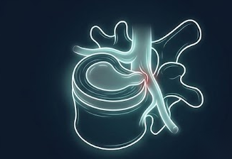
A herniated disc can pinch a nerve to the leg

A catch-all term for the age-related wear, bulging, and herniation of the cushioning discs in the lower back, which can crowd the nerves that travel to the legs.
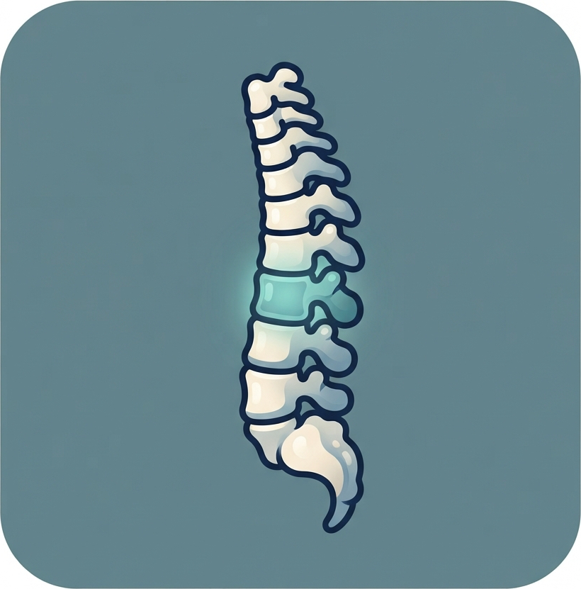
Between each pair of lower-back bones sits a disc: a tough outer ring around a soft, gel-like center. With age these discs lose water and height and the outer ring weakens. Sometimes the soft center pushes through the ring — a herniation — into the spinal canal, where it can press on a nerve root. When that happens, the classic result is sciatica: pain, numbness, tingling, or weakness that travels from the low back or buttock down a specific path into the leg and foot.
Which nerve is affected depends not only on the level but on the direction of the herniation. A herniation just off the midline (paracentral) usually catches the nerve traveling past that level (the traversing root), while a herniation out to the side (far-lateral) tends to catch the nerve exiting at that level. You can explore this in the interactive tool below.
The good news is that most lumbar disc herniations improve on their own over weeks to months. When surgery is needed, our approach at Brown is least-invasive-first: relieve the nerve with the smallest effective operation, and reserve fusion for situations where the spine is genuinely unstable. The reasoning behind each option is explained throughout this page.
The soft disc center pushes through the outer ring into the canal and can press on a nerve root to the leg.
A pinched nerve sends pain, numbness, or weakness down a specific path into the buttock, leg, and foot.
A paracentral herniation hits the traversing root; a far-lateral one hits the exiting root at that level.
Most herniations shrink and settle over weeks to months, so patience and non-surgical care come first.
Most sciatica is not an emergency, but seek care right away for: new loss of bladder or bowel control, numbness in the groin or inner thighs (the "saddle" area), or rapidly worsening weakness in one or both legs. These can signal cauda equina syndrome — severe compression of the nerve bundle — which is a surgical emergency.
Relieving pressure on a nerve is very good at easing leg symptoms and less reliable for back pain alone. Select a symptom to see what to realistically expect.
Select a symptom to see how treatment typically affects it.
Different lumbar levels and herniation directions map to different nerves, which is why one person feels the top of the foot and another the sole. Pick a level and herniation direction to see which nerve root that pattern most often fits — educational, not a diagnosis.
This interactive tool is provided by the Norman Prince Spine Institute. It is a pattern-recognition aid for education and should be correlated with your exam and imaging by a clinician.
Several factors shape whether surgery makes sense and which operation fits. Choose a category, then select a factor to learn more.
Select a factor to see how it affects candidacy and which operation fits.
Most people improve without surgery. An operation is considered when leg symptoms are severe or persistent despite a fair trial, when there is significant weakness, or urgently if there are signs of cauda equina syndrome.
The majority of lumbar disc herniations improve within six to twelve weeks. Physical therapy, activity changes, anti-inflammatory or nerve-pain medication, and, in selected cases, an epidural steroid injection can control symptoms while the herniation shrinks and the nerve settles. Surgery is rarely urgent unless there is major weakness or cauda equina syndrome.
When surgery is warranted, the goal is simply to remove the fragment of disc pressing on the nerve — not the whole disc, and usually without fusing anything. This can be done through a small open incision (microdiscectomy) or an even smaller endoscopic portal. Fusion is added only when the segment is also unstable.
Select an operation to see, in one place, how it works step by step and what to expect. At Brown we favor the least-invasive effective approach and reserve fusion for genuine instability.
Endoscopic surgery is a signature strength of our division. Explore our interactive tool to see just how small the portal is compared with other approaches, and what the surgeon sees through the scope as the fragment is removed.
When a disc problem also needs fusion, the level is held with screws and rods. See how those screws are placed through incisions about a centimeter long, using robotic navigation.
All three operations, side by side. For a straightforward herniation the first two both remove the offending fragment and free the nerve — the difference is mostly the size of the corridor; fusion is added only for genuine instability. The best choice depends on your anatomy and the herniation, decided with your surgeon.
| Operation | Corridor | Fusion | Best for | Typical recovery |
|---|---|---|---|---|
| MicrodiscectomySmall open incision · microscope | Small incision (~2–3 cm), microscope | No — motion preserved | Most herniations, including larger or migrated fragments | Usually same day; light activity in days to ~2 weeks |
| EndoscopicPencil-width portal · camera | Pencil-width portal, high-definition scope | No — motion preserved | Focal herniations in favorable positions | Usually same day; return to light activity in days |
| Discectomy + FusionDecompress and stabilize | Fragment removed, then a cage and screws | Yes — the level is joined | Herniation with instability or slippage | 1–2 nights; longer while the fusion heals |
None is universally better. The aim is to remove exactly what is pinching the nerve through the smallest corridor that does the job safely, and to add fusion only when the level is genuinely unstable.
This educational resource was developed by the Division of Spine and Spine Tumor Neurosurgery at Brown University Health to help patients and families understand lumbar disc disease and its treatment options. Our surgeons offer the full spectrum of care, from endoscopic and minimally invasive discectomy to fusion when needed, and work closely with physical therapy, pain management, and physiatry so that surgery is recommended only when it is the right next step.


For full faculty profiles, visit the Brown Neurosurgery Spinal Surgery Division.