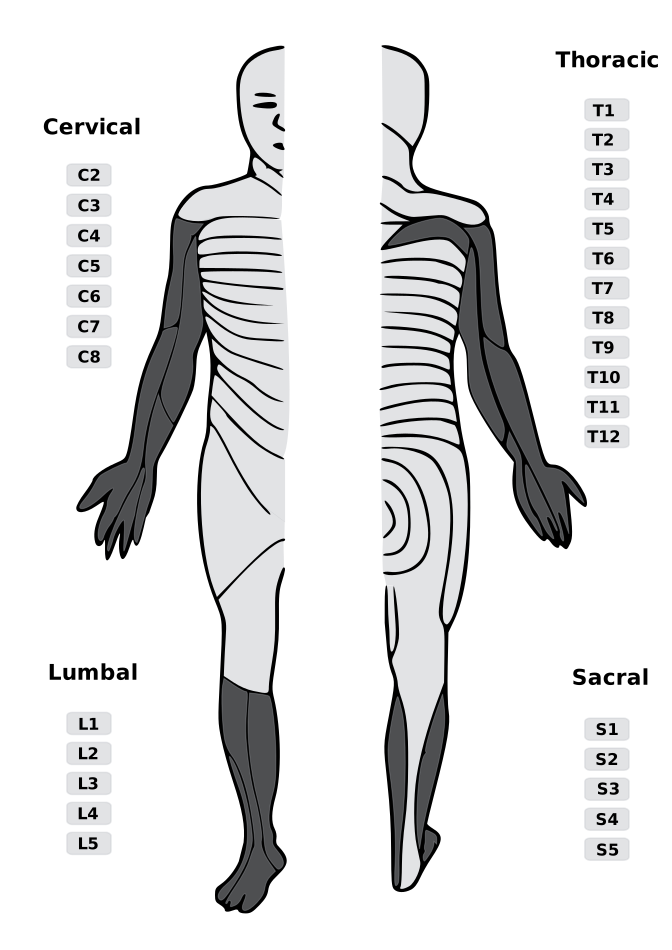
Cervical · Upper-extremity pattern aid
Cervical disc level → exiting root.
In the cervical spine, disc-level naming maps differently than lumbar levels: a C5–C6 disc lesion most classically affects the C6 root, a C6–C7 lesion affects C7, and C7–T1 affects C8.
Selected cervical level
C6–C7
Affected root
C7
exiting cervical root
Dermatome (sensory)
Myotome (motor)
Reflex
Bedside clue
Dermatomal map
C7 highlighted
Cervical upper-extremity zones
| Disc level | Classic root | Dermatome | Motor | Reflex | Common mimic / caution |
|---|
Cervical clinical warning
- C7 is often reported as the most frequently involved cervical nerve root, while C2–C4 radiculopathy is uncommon [Kang et al.].
- Classic patterns are imperfect: McAnany et al. found standard textbook pain/numbness patterns in only 54% of 239 single-level surgical cases, with nonstandard patterns differing by an average of 1.68 dermatomal levels [McAnany et al.].
- Screen for myelopathy when symptoms include hand clumsiness, gait imbalance, hyperreflexia, Babinski, clonus, Hoffmann sign, or bowel/bladder change; signs or symptoms of myelopathy warrant urgent referral [McCartney et al.].
Cervical root patterns synthesize Kang et al., McCartney et al., and the Merck Manual.