What Is It? Symptoms Watch or Treat? How It's Treated Request Consultation
Division of Neurotrauma
Interactive Patient Education

Chronic Subdural Hematoma
...and What to Do About It

A chronic subdural hematoma is a slow, quiet collection of blood that builds between the brain's surface and its outer covering over the course of weeks or months often starting with a fall or bump too minor to remember. Because it builds so gradually, it can be mistaken for a stroke, dementia, or ordinary aging. Treatments range from observation to surgery to endovascular embolization.

What Is a Chronic Subdural Hematoma?

A slow bleed between the brain and its outer covering that builds up over weeks or months.

A subdural hematoma is a collection of blood over the surface of the brain (not within the brain). A chronic subdural hematoma forms when the rate of reabsorption doesn't keep up with the rate of blood leakage, so there can be a gradual build up of fluid and pressure on the brain over weeks to months.

A fall or bump, even one minor enough to be forgotten entirely, can tear a brain surface vein and cause it to leak. Unlike an acute subdural hematoma, a large, sudden clot that follows a severe head injury and is usually a same-day emergency, a chronic one accumulates slowly, and may cause gradual symptoms such as headache or imbalance.

Not every chronic subdural hematoma needs treatment right away. A small, mild collection is often simply watched with a follow-up scan. When treatment is needed, it may involve neurosurgery, such as burr hole drainage or a craniotomy, an endovascular procedure called middle meningeal artery embolization, or a combination of the two, depending on the size and internal makeup of the blood collection.

Key Facts
Type: A slow collection of blood between the brain's surface and the dura, its outer covering
Who's typically affected: Older adults and people on blood thinners, most often after a minor or forgotten fall
Diagnosis: CT or MRI of the head
Builds over: Weeks to months after the triggering injury
Treatment range: Careful observation, surgical drainage, or endovascular embolization of the leaky vessel.

Use the controls below to grow and age a subdural collection. Observe how the brain shifts in response to a growing collection, and how the internal structure changes the longer the subdural hematoma persists.

Size & Age of Subdural Hematoma
Collection Size 0% · Age 0%
Age of Subdural Hematoma →
Click and drag this point
Size of Subdural Hematoma →
Location
Reference Presets
Key Features & Significance
Normal brain, no extra-axial collection.

Which Symptoms Point to a Chronic Subdural Hematoma?

Because the bleed builds slowly, its symptoms often look like something else entirely. Select an item to learn more.

Sudden, severe symptoms after a fall are still an emergency

A chronic subdural hematoma can occasionally bleed acutely on top of its slow baseline. Any sudden, severe headache, new one-sided weakness, difficulty waking up, or loss of consciousness after a fall or head injury, at any age, needs immediate emergency evaluation. Do not wait to see if it improves.

Select an item on the left to learn more.
The Root Cause: Why Does a Chronic Subdural Hematoma Form?

In many cases, the story starts with a set of small, stretched veins on the surface of the brain.

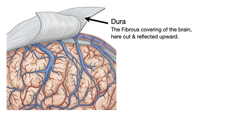
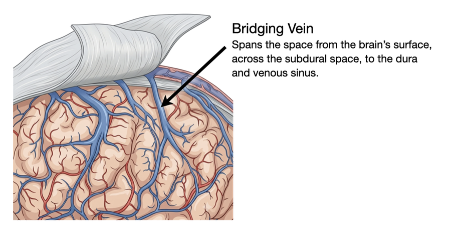
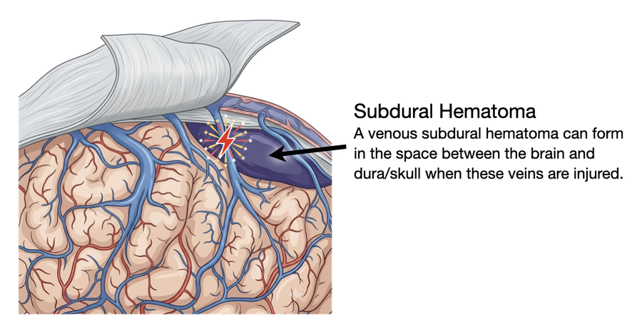
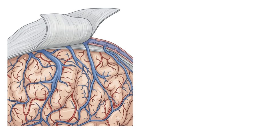
The brain sits inside the skull wrapped in the dura mater, a tough, fibrous membrane. Between the brain's own surface and the dura is a thin, fluid-filled space, and running across that gap are a handful of small cortical veins, "bridging veins," that carry blood from the brain's surface into the large draining sinuses built into the dura itself.

As the brain gradually shrinks with age — a normal process that speeds up with conditions like chronic alcohol use — the space the veins must cross widens. The bridging veins stretch further to make the same connection, leaving them thinner, tauter, and more fragile than they were years earlier.

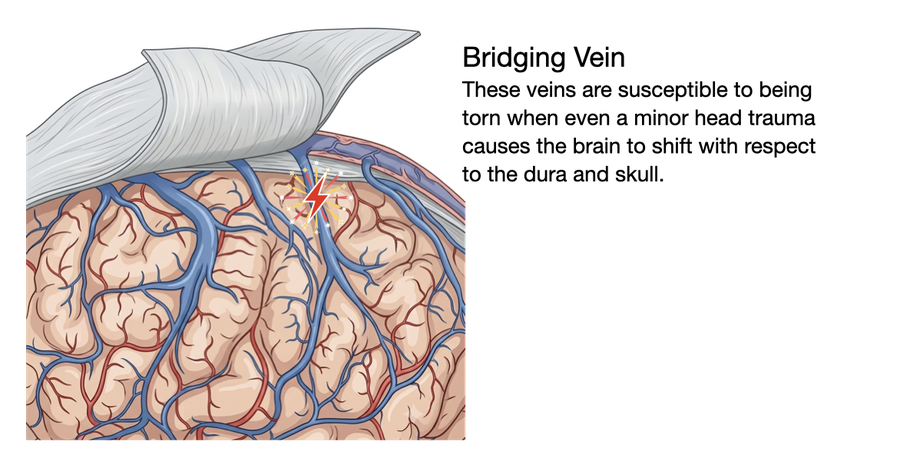
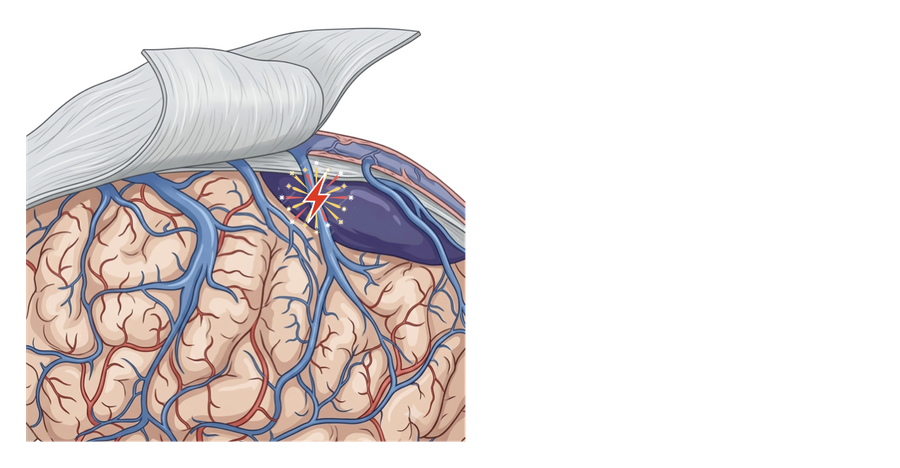
In many, though not all, cases, that's where a chronic subdural hematoma begins: a jolt or a fall, often minor enough to be forgotten by the time symptoms appear weeks or even months later, shears one of these stretched veins right where it crosses into the sinus. Because bridging veins carry low-pressure venous blood rather than the higher-pressure blood of an artery, the resulting leak is slow, which is why the blood collects gradually rather than causing an immediate, obvious neurologic change.

Diagram of the brain's surface beneath the dura, showing cortical bridging veins crossing the subdural space to drain into a dural venous sinus. The same diagram with an arrow highlighting one bridging vein as it crosses the subdural space toward the sinus. A closer view of that bridging vein at the exact point where it crosses into the dural sinus. The bridging vein shearing at the point where it crosses into the sinus. Blood beginning to collect in the subdural space at the site of the torn vein. A larger pool of blood in the subdural space, marking the start of a chronic subdural hematoma.

Not the only path to a subdural hematoma

Bridging vein shearing is the most common cause, especially in older adults and people taking blood thinners, but it isn't the only one. A subdural hematoma can also form from direct injury to a small artery or vein on the brain's surface, from an underlying bleeding disorder, from the slow enlargement of an earlier acute bleed or, occasionally, with no clear preceding injury at all.

Whether, and How to Treat?

There is rarely one single right answer. Several factors together guide whether we recommend observation, drainage, embolization, or a combination. Select a factor to learn how it weighs in.

Select a factor on the left to learn more.

Burr Hole Drainage, Craniotomy, or Middle Meningeal Artery Embolization

These three approaches aren't strictly either/or, and which one (or which combination) is right depends heavily on what the collection looks like on imaging: whether it's a single pocket of fluid, divided by membranes, or partly solid. Many patients, especially with a larger, recurrent, or more complex collection, are offered more than one.

Burr Hole DrainageCraniotomyMiddle Meningeal Artery Embolization
What happensOne or two small, nickel-sized holes are drilled through the skull directly over the collection, and the old blood is drained through a thin catheter.A larger, though still limited and temporary, section of skull is opened so the surgeon can see directly into the space, open any internal pseudomembranes, and remove both liquid and more solid, subacute clot.A thin catheter is guided from an artery in the wrist or groin up to the middle meningeal artery, the vessel feeding the abnormal membrane around the collection, which is then intentionally blocked from the inside.
AccessThrough the skull, directly over the collectionThrough the skull, using a larger temporary bone opening that is replaced at the end of surgeryEntirely through the blood vessels; no skull opening at all
What it treatsRemoves the fluid that has already collected, relieving pressure right awayRemoves fluid, internal membranes, and solid or subacute clot that a burr hole and catheter cannot adequately reachDoes not remove existing fluid on its own; it starves the membrane's abnormal blood supply so the collection is less likely to keep growing or return
Typical hospital stay1–3 days, with a drain often left in for 24–48 hoursTypically 3–5 days, reflecting the larger opening and more extensive procedure1 additional day if combined with surgery, or a short stay if used alone

Why a Temporary Drain Is Often Left in Place

After either burr hole drainage or craniotomy, a thin, soft subdural drain is often left in the space where the collection was for up to a few days afterward. Removing the fluid doesn't immediately close the space it occupied, and that space can slowly refill before the brain has had a chance to re-expand and settle against the skull. A temporary drain lets any fluid that continues to seep in keep draining externally during this early window instead of building back up, which meaningfully lowers the chance of recurrence. It's typically removed at the bedside once imaging or drainage output shows it's no longer needed, before you go home.

How It's Treated, Step by Step

From diagnosis to follow-up, the path can branch depending on what's right for you. Select any step below to see what happens and why.

Select a step to learn what happens — and why.

What Outcomes Can Patients Expect?

Evidence from clinical series and randomized trials, presented honestly, including the risks.

80–90%
of patients have meaningful improvement in symptoms after drainage of a chronic subdural hematoma.
10–20%
chance the collection recurs after burr hole drainage alone (up to about a third in some series), which is why good follow-up, possibly with more imaging, is important.
~50%
relative reduction in that recurrence risk when middle meningeal artery embolization is added, based on pooled randomized-trial data.

Risks and Side Effects

The most common complication is recurrence, fluid building back up and requiring a repeat procedure, which happens in roughly 10–20% of patients after drainage alone. Seizures can occur in the days to weeks after surgery. Bleeding or infection at the drainage site is uncommon but possible. Craniotomy involves a larger opening than burr hole drainage and carries a modestly higher risk of bleeding or infection, though still low; it is generally reserved for the more complex collections described above, where a burr hole would not adequately drain the membranes or solid clot present. Anesthesia carries its own risks, which are magnified in frail, older patients, and are weighed carefully when planning surgery for this group.

For embolization specifically, risks relate mainly to the catheter access site (bruising, rarely a vessel injury) and, uncommonly, unintended blockage of a nearby vessel. Pooled trial data put the overall complication rate from the embolization procedure itself at around 1%.

Common Questions

I don't remember hitting my head. How could I have this?
Many patients, especially older adults or those on blood thinners, don't recall the fall or bump that caused it. The injury is often minor enough at the time to be forgotten entirely, and the bleeding itself builds slowly over weeks rather than causing an immediate problem.
Do I need surgery right away, or can we watch it first?
It depends on the size of the collection and whether it's causing symptoms. Small, incidentally found collections with no or minimal symptoms are often safely observed with a repeat scan in a few weeks. Larger or clearly symptomatic collections are usually treated surgically, with the specific approach (burr hole drainage or craniotomy) chosen based on what the collection looks like on imaging.
What is burr hole surgery like?
One or two small, nickel-sized holes are drilled through the skull directly over the collection, and the old blood is drained through a thin tube. A drain is often left in place for a day or two afterward to let any remaining fluid continue to clear.
Will I need burr holes, or something bigger like a craniotomy?
Most collections are a single, continuous pocket of fluid and are well suited to burr hole drainage. But imaging sometimes shows plentiful pseudomembranes, internal walls dividing the collection into several separate compartments, or a more solid, subacute portion of partly clotted blood mixed in with the liquid part. Either finding means a small burr hole and thin catheter likely won't clear the whole collection, so a craniotomy, a somewhat larger but still temporary opening, is planned instead so the membranes can be opened and every part of the collection, liquid and solid, removed under direct vision. Your surgeon will go over which approach fits your imaging before surgery.
What is embolization, and do I need it in addition to surgery?
It's a minimally invasive, catheter-based procedure that blocks the artery feeding the membrane around the collection, to help keep it from coming back. It isn't needed for every patient; it's one of the main options especially for a collection that has already recurred, or when paired with burr hole drainage or craniotomy for a larger or more complex one.
Can this come back after treatment?
Yes. Recurrence is the most common complication, happening in roughly 10–20% of patients after drainage alone, which is why follow-up imaging is routine. Adding embolization roughly cuts that risk in half.
Should I stop taking my blood thinners?
Never stop a blood thinner on your own. Your neurosurgery team and prescribing physicians will weigh the bleeding risk from the hematoma against the original reason you're on the medication, such as a heart valve or a history of blood clots, and give you a specific plan.
Is this the same as a stroke?
No, though it can look similar from the outside. A stroke is a sudden loss of blood flow to part of the brain; a chronic subdural hematoma is a slow buildup of blood on the brain's surface. Imaging tells the two apart immediately, which is why anyone with new one-sided weakness should be evaluated urgently regardless of the suspected cause.
How long until I feel like myself again?
Many patients notice improvement within days of drainage, though full recovery of thinking and walking can take several weeks, especially in older patients. Your team may schedule follow-up imaging to confirm the collection isn't returning.

Meet the Team

Chronic subdural hematoma is managed within Brown Neurosurgery's Neurotrauma division, in partnership with our Endovascular Neurosurgery Program for cases suited to embolization.

What Is It? Symptoms Root Cause Watch or Treat? Treatment Options How It's Treated Outcomes FAQ