What Is This? Living With a Skull Defect Timing and Type of Cranioplasty Request Consultation
Division of Neurotrauma
Interactive Patient Education

Understanding
Craniectomy
and Cranioplasty

After a severe brain injury, stroke, or bleed, surgeons sometimes remove a section of skull so the brain has room to swell safely. This page walks through why that happens, what it's like to live with a skull defect in the meantime, and the later surgery, cranioplasty, that reconstructs it.

What Is Craniectomy? What Is Cranioplasty?

Two different operations, done at two different times, for two different reasons.

The skull is a rigid, closed box. Under normal conditions that's a good thing: it protects the brain. But when the brain swells, or when blood collects inside the skull after an injury, stroke, or hemorrhage, that same rigid box becomes a problem: there is nowhere for the extra volume to go, so pressure inside the skull (intracranial pressure) climbs. If it climbs high enough for long enough, the brain's own blood supply can be choked off, causing further, often irreversible, damage.

A craniectomy removes a section of skull bone and, unlike a routine craniotomy, does not put it back right away. That missing piece gives the swollen or bleeding brain somewhere to expand that isn't inward against itself. It is a temporary, deliberately incomplete operation: the goal in the moment is survival and protecting brain tissue, not a finished result.

Cranioplasty is the second operation that completes the story: once the swelling has resolved and the patient has recovered, surgeons reconstruct the missing section of skull, using either the patient's own stored bone flap or a custom synthetic implant. Brown's Neuroplastic Center brings neurosurgeons and plastic and reconstructive surgeons together specifically for this decision, since the best material and timing choice is rarely obvious and often benefits from more than one specialist's perspective.

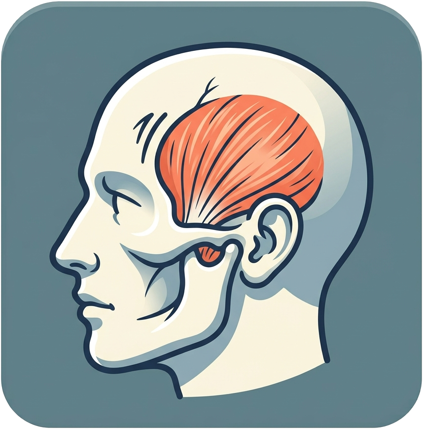
Illustration of a skull in profile opened to reveal the brain, representing craniectomy
Key Facts
Craniectomy timing: Usually urgent or emergency
Cranioplasty timing: Planned, typically weeks to a few months later
Materials: Patient's own bone, or titanium, PMMA, or PEEK implants
Anesthesia: General anesthesia for both operations

When Is Craniectomy Needed?

Select a cause to see how it can lead to dangerously high pressure inside the skull.

From Injury to Craniectomy to Cranioplasty

The Care Journey

Select a step to learn what happens and why.

Syndrome of the Trephined

When part of the skull has been removed to relieve pressure on the brain, that part of the brain is protected only by skin. In some cases, over time, ordinary atmospheric pressure can push the flap inward, causing symptoms. Learn here about specific symptoms that may arise in certain cases.

The Severe End of the Spectrum: Paradoxical Herniation

In rare, severe cases, the pressure gradient across the defect becomes strong enough that the brain itself shifts inward through the opening, called paradoxical herniation. This can cause rapid neurological decline and is treated as an emergency. It's part of why the team watches for syndrome of the trephined closely rather than waiting for it to become severe.

Choosing a Cranioplasty Material

There is no single "best" material for every patient. Select an option to see how it compares.

When Is the Right Time for Cranioplasty?

Timing is individualized, but tends to fall into three general windows.

Ultra-Early · Within ~5 Weeks

Faster Return to Normal Life

May allow earlier relief from syndrome of the trephined symptoms and a quicker return to normal activity, but carries a somewhat higher risk of fluid buildup (hydrocephalus) if the brain hasn't fully settled.

Early · Roughly 5–12 Weeks

The Window Most Surgeons Favor

Swelling has usually resolved and the scalp has fully healed, without waiting so long that the soft tissue has tightened firmly over the open defect.

Late · Beyond ~12 Weeks

Sometimes Necessary

Infection, wound healing problems, or other medical issues can delay surgery. Timing is always adjusted around what's safest for that patient, not a fixed calendar date.

Timing and Type of Cranioplasty

Several factors shape when cranioplasty happens and which material is used. Select one to learn more.

Temporal Hollowing and Muscle Wasting

A sunken temple is not the same problem as a skull defect, and fixing the bone alone does not fix it.

Illustration of a head in profile with the fan-shaped temporalis muscle at the temple highlighted, the muscle affected by temporal hollowing

The temporalis muscle is a fan-shaped chewing muscle that sits just under the skin at the temple, directly over the bone that many cranial approaches need to reach. During surgery, it is often detached, retracted, or thinned to expose the skull, and it does not always heal back to its original bulk or position. The result, in a large share of patients after this kind of approach, is a visible flattening or dent at the temple, called temporal hollowing, that persists even after cranioplasty has fully reconstructed the bone underneath it.

Because this is a muscle and soft-tissue problem rather than a bone problem, Brown's Neuroplastic Center approaches it as a true team decision between neurosurgery and plastic and reconstructive surgery, matching several dedicated techniques to how mild or pronounced the hollowing is.

Minimally Invasive

Fat Grafting

The patient's own fat is harvested from elsewhere in the body and injected to fill mild-to-moderate hollowing. It's a relatively minor procedure, though results can soften somewhat over time and occasionally need a touch-up.

Restores Anatomy

Muscle Repositioning

The temporalis muscle is surgically returned to its natural position, often combined with cranioplasty in the same operation. This addresses the underlying cause rather than just adding volume on top of it.

For Significant Hollowing

Custom Implant Augmentation

A shaped implant, sometimes built directly into the cranioplasty implant itself, restores lost temple volume. This is often the most reliable option for more pronounced or long-standing hollowing.

What Outcomes Can Patients Expect?

Both operations are generally well tolerated, but all neurosurgical procedures carry risks.

Fewer Deaths
Timely hemicraniectomy for large strokes has been shown in clinical trials to significantly reduce the chance of dying, though many survivors still live with meaningful disability, a trade-off patients and families weigh directly with their team.
~5–8%
Approximate infection risk after cranioplasty, one of the most common reasons for a repeat operation.
Resorption Risk
Autologous bone can gradually resorb over months to years, more often in children and fragmented flaps, sometimes needing a second cranioplasty with a synthetic implant.

Craniectomy Risks

  • Bleeding or infection at the surgical site
  • The underlying risks of the emergency itself (stroke, injury, or hemorrhage)
  • A vulnerable period requiring a protective helmet
  • Syndrome of the trephined while the defect remains open

Cranioplasty Risks

  • Infection (roughly 5–8%)
  • Bone resorption (autologous bone specifically)
  • Hardware or implant exposure, bleeding, or hematoma
  • Seizure or hydrocephalus, especially with very early timing

Common Questions

Will I need to wear a helmet the whole time?
Generally, yes, whenever you're up and moving around, until cranioplasty is performed. With the bone flap out, that part of the brain has no bony protection at all, so even a fairly minor bump could be dangerous without it.
How long will I have the skull defect before cranioplasty?
It's individualized, typically somewhere from a few weeks to a few months. The team waits until swelling has resolved and the scalp has fully healed before scheduling the reconstruction, since operating too early or too late each carries its own tradeoffs.
Can I use my own bone, or do I need an implant?
It depends on whether your original bone was stored properly and remains healthy, how the defect is shaped, and your own preferences. The team discusses both autologous bone and synthetic options directly with you rather than defaulting to one.
Will cranioplasty fix the sunken look at my temple?
Not by itself. The skull defect and temporal hollowing are two different problems: one is missing bone, the other is a muscle that didn't heal back to its original shape. Cranioplasty addresses the bone; fat grafting, muscle repositioning, or a custom implant address the temple, and can often be done in the same operation.
Is craniectomy reversible?
Yes. Unlike many neurosurgical treatments, a craniectomy is explicitly meant to be temporary, and cranioplasty is the planned second step that completes the reconstruction.
What if my symptoms feel worse when I sit up?
That pattern, feeling worse upright and better lying flat, is a classic sign of syndrome of the trephined. It's worth reporting to your team promptly, since it can influence how soon cranioplasty should be scheduled.
How risky is the cranioplasty surgery itself?
Most patients tolerate it well, recover within a few weeks, and many notice real improvement in headache, fatigue, or thinking that appeared while the defect was open. That said, it is a real operation with real risks, including infection and, for autologous bone, resorption over time, which your surgical team will walk through with you individually.

Meet the Team

Craniectomy and cranioplasty care at Brown Neurosurgery draws on both neurotrauma neurosurgery and the Neuroplastic Center's plastic and reconstructive surgery expertise.

What Is This? When Is It Needed? The Care Journey Syndrome of the Trephined Choosing a Material Timing and Type of Cranioplasty Temporal Hollowing Outcomes FAQ