What Is This? Warning Signs Treatment Recovery Request Consultation
Division of Neurotrauma
Interactive Patient Education

Traumatic Brain Hemorrhage:
Types and Treatments

A blow to the head can cause bleeding in or around the brain in several different ways. Different types of bleeding can raise different concerns and require specific management strategies. This page walks through the different factors that may come into play and how Neurotrauma and Neurocritical Care teams may respond.

For any immediate concerns regarding a head injury, call 911 or go to the nearest emergency department now

This resource should not be used to make decisions regarding acute care. The information on this page is intended to inform patients and family members about the basic types traumatic intracranial hemorrhage and the management decisions that clinicians may need to make when interacting directly with patients who present with traumatic brain injury.

What Is a Traumatic Intracranial Hemorrhage?

Bleeding inside the skull after an injury is not one condition. It is several different conditions that happen to share a cause.

A traumatic intracranial hemorrhage is bleeding inside the skull caused by an injury: a fall, a motor vehicle collision, a sports impact, an assault, or any other blow or sudden movement of the head. It is different from a concussion, where imaging typically looks normal even though something has clearly happened to how the brain is working. When a scan does show blood, the single most useful question a clinician asks is not just "how much blood," but "where is the blood." Location determines the likely cause, what the bleed will look like on a CT scan, how quickly it is likely to change, and how urgently it needs treatment.

To understand why location matters so much, it helps to picture the layers between the outside of the head and the brain itself: the skull, then two layers of tough membrane called the dura mater, then a thinner membrane called the arachnoid, a fluid-filled space, and finally the surface of the brain itself, which has an even thinner membrane (the pia) hugging it directly. These layers create "compartments" that define how blood may expand, spread, and cause injury to the brain itself. The next section walks through each of these compartments and the type of bleed that occurs in it.

Key Facts
Common causes: Falls (the single leading cause), motor vehicle collisions, sports and recreational impacts, and assaults
First-line imaging: CT scan, usually within minutes of arrival
Urgency: Ranges widely, from close observation to emergency surgery within the hour
The core idea: The skull is a closed, rigid box — location and pressure effects matter as much as how much blood there is

Not All Brain Bleeds Are the Same

Select a type below to see where it occurs, why it happens, and what it typically means for treatment and outlook. The same slider also controls the pressure graph on the right, since not every type of bleed pushes intracranial pressure the same way.

Total Skull Volume (fixed)
Brain tissue CSF & blood (displaceable buffer) Added blood / swelling
Added Volume → Pressure (ICP) →
The intracranial pressure–volume curve: flat at first, then steep once the brain runs out of room to compensate.
Select a type on the left to see where it occurs.
Why Small Bleeds Can Become Emergencies

The skull is a closed, rigid container. Blood takes up space at the expense of other skull contents, including the brain itself. This is known as the Monro-Kellie doctrine. At some point, the ability to compensate for the added volume of blood (by decreasing other fluids, including cerebrospinal fluid, for example) diminishes, and pressure starts to rise more quickly. Drag the slider above to see what happens to intracranial pressure as the volume of blood, and any associated swelling, increase — and notice how differently each type of bleed moves along this same curve.

Normal — compensating well At this stage, the brain compensates by shifting CSF and venous blood out of the way, so pressure inside the skull stays close to normal.

What Symptoms Mean What

Not every symptom after a bump on the head means a dangerous bleed, and not every dangerous bleed causes symptoms right away. Select a symptom below to see how it is generally regarded.

Select a symptom on the left to learn more.

Diagnosis and Monitoring

A combination of the neurological exam, brain imaging, and sometimes invasive monitors, allows us to follow a patient’s status.

Assessing Level of Consciousness: the Glasgow Coma Scale

The Glasgow Coma Scale (GCS) is a tool that was designed to assess the initial severity of brain injury by allowing medics, doctors and nurses to quickly rate a person’s level of awareness and function in the field or at the bedside. In the emergency department, assessment of GCS helps assign patients to the proper TBI protocol, such as whether intracranial pressure monitoring is indicated. The GCS may be used to track changes over time, but a more specific neurological exam is usually preferred once a patient is settled in the hospital.

Eye Opening

Up to 4 points

Verbal Response

Up to 5 points

Motor Response

Up to 6 points
GCS 13–15Mild
GCS 9–12Moderate
GCS 8 or belowSevere
Total Score
Select a response in each category above

Neuroimaging

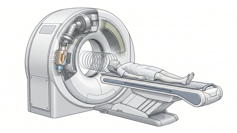
Illustration of a CT scanner with a patient positioned for a head scan

CT Scan — First and Fastest

A CT scan is almost always the first imaging study after a significant head injury: it is fast, widely available, and shows blood clearly. We look at the type of bleed, its size, whether it is pressing on or shifting the brain, and whether the fluid spaces (ventricles) that normally cushion the brain are being compressed. Repeat CT scans at a set interval are often routine, not necessarily a sign that something has gone wrong. Subdural and epidural hematomas can continue to expand over time, and CT scans are often used to track this, and to assess whether surgery may be required. Contusions, meanwhile, can blossom (enlarge) over the first hours to days, and a repeat scan catches that early, before it shows up as a change in how a patient is behaving.

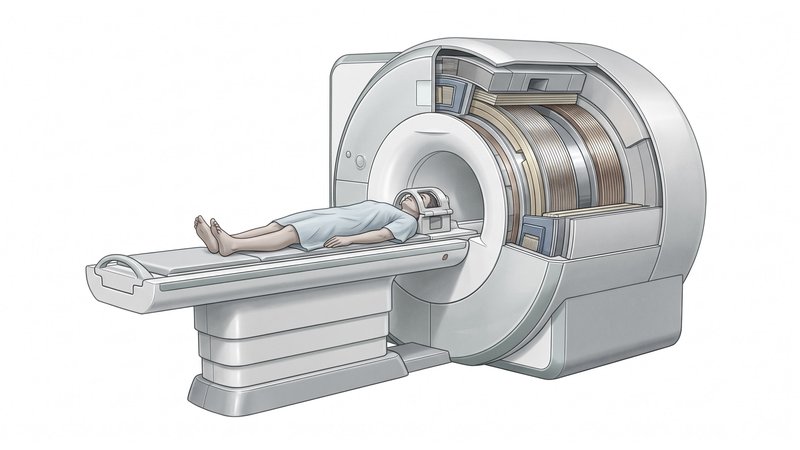
Illustration of an MRI scanner with a patient positioned for a head scan

MRI — More Sensitive, Used Later

MRI is not typically the first study in the emergency setting; it takes longer and is harder to perform safely in an unstable patient. Once someone is stable, however, MRI — especially sequences sensitive to blood breakdown products — is considerably more sensitive than CT for diffuse axonal injury, which can look deceptively normal on an initial CT scan despite causing real symptoms. In addition, MRI does a better job showing the brain itself and swelling due to nearby blood. Overall, MRI may be a better indicator of the extent of injury, and may help us determine a patient’s prognosis.

Intracranial Pressure (ICP) Monitoring

For more severe injuries, imaging and the GCS are not enough on their own — we sometimes place a dedicated pressure monitor directly inside the skull so intracranial pressure (ICP) can be measured continuously in real time, rather than inferred indirectly. This connects directly to the idea explored earlier: once pressure can be measured moment to moment, it can be treated before it causes permanent damage rather than after. There are two main ways this is done.

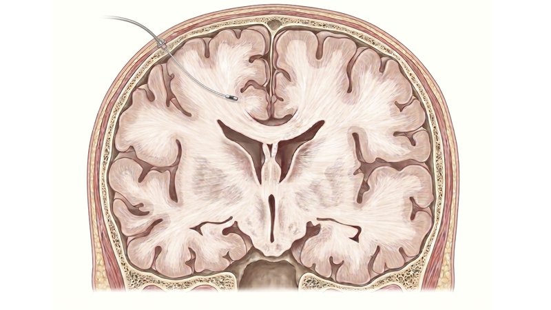
Illustration of an intraparenchymal ICP monitor (bolt) placed a few centimeters into brain tissue

Intraparenchymal Monitor (“Bolt”)

Monitors pressure

A thin pressure-sensing probe is passed through a small hole (“bolt”) in the skull and a few centimeters into the brain tissue itself. It is quick to place, does not require finding a specific fluid space, and gives a continuous, reliable pressure number at the bedside. Its limitation is that it only measures pressure — it cannot be used to drain fluid the way an EVD can.

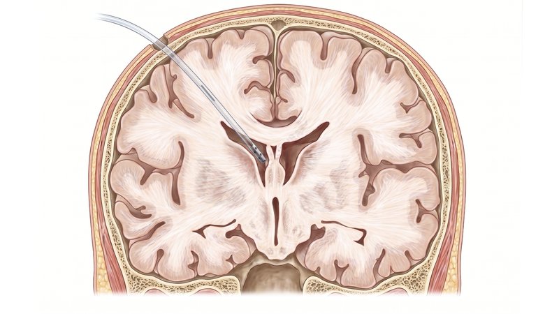
Illustration of an external ventricular drain (EVD) catheter placed into a brain ventricle

External Ventricular Drain (EVD)

Monitors and treats pressure

A thin catheter is guided into one of the brain’s fluid-filled ventricles and connected to an external monitor. Beyond measuring pressure, an EVD can also actively drain cerebrospinal fluid (or blood, in the case of IVH) to directly relieve pressure — making it both a monitoring tool and a treatment, which is why it is often preferred when the ventricles are enlarged or when IVH is present.

Watching the Waveform

An ICP monitor reports the average pressure in the brain, and also provides a continuous pressure waveform, one pulse per heartbeat. The shape of that waveform carries information about the brain’s compensatory reserve (its “compliance” or ability to compensate for increased mass effect from bleeding or swelling), often before the average pressure itself looks concerning on its own. Learn about the ICP waveform below.

8 mmHg

As reserve is used up, watch P2 rise to meet, then exceed, P1 — and notice how the two graphs move together.

normal ≤2 pathological >4 Intracranial Pressure (0–30 mmHg) → Elastance (mmHg/mL) →
Elastance (pressure rise per mL added) climbs quickly as soon as reserve starts to decline, then rises more gradually as ICP itself continues to climb . The dashed lines mark the normal (≤2) and pathological (>4 mmHg/mL) thresholds.
Normal — clean, well-defined waveform P1 is clearly taller than P2, with sharp, distinct peaks and low pulse amplitude.

When Surgery Is Needed Right Away

Some bleeds are so large that they press on the brain to compromise blood-flow and function. When that hematoma is large enough, or the patient is declining, removing it is the most direct way to relieve pressure.

Epidural Hematoma

Because these are often arterial and can expand quickly, all but the smallest are typically evacuated through a craniotomy promptly, and even small ones are watched closely in the ICU rather than sent home.

Acute Subdural Hematoma

Often evacuated when it is causing significant "mass effect" or neurological decline; a small one in a patient who is neurologically stable may instead be watched closely with repeat imaging. A slower-forming, older version of this same condition is covered separately on our Chronic Subdural Hematoma page.

Large Contusion

Contusions that have "blossomed" enough to cause severe mass effect or swelling may require a decompressive craniectomy to relieve pressure on the brain.

The "Lucid Interval"

Epidural hematomas classically feature a lucid interval: because the blood has not yet touched the brain directly, a patient can seem entirely normal for a stretch of time before rapidly declining as the bleed expands and the pressure begins to affect neurological function. This is why even a small, asymptomatic epidural hematoma is watched closely rather than dismissed.

A Tiered, Escalating Approach

When there is no single mass to remove, or after one has been removed, treatment shifts to directly controlling pressure inside the skull. Select each tier to see what it involves and why it comes where it does.

Interactive · How Hyperosmolar Therapy Works
Why Mannitol and Hypertonic Saline Pull Water Out of the Brain
A “tier 2” intervention for heightened intracranial pressure is giving hyperosmolar therapy, such as mannitol or a concentrated salt solution. These work by shifting water across the blood–brain barrier. Take a deeper dive here to see how these treatments work.
Try the water-shift model
Brain Tissueinterstitial space
Blood–Brain
Barrier
Blood Vesselcapillary lumen
87%
BrainWaterConcentration
87%
VesselWaterConcentration
Water molecule Solute (mannitol / Na⁺Cl⁻) Net Water Flow

Water moves toward whichever side has proportionally less free water because random molecular motion, over time, evens out the water fraction on both sides of a membrane that only water can cross. Giving a hypertonic IV solution (like mannitol or 3% saline) raises the solute concentration in the blood vessel; water then moves from the (relatively more dilute) brain tissue into the vessel until the two sides re-equilibrate. That water, no longer in the swollen brain, ultimately leaves the body in urine, visibly shrinking brain tissue as it leaves, counteracting swelling.

What Outcomes Can Patients Expect?

Outcomes vary enormously depending on the type of bleed, its severity, and a patient’s age and overall health. Even knowing these things, precisely predicting long-term outcomes for any particular individual with traumatic brain injury is still fraught with difficulty and uncertainty.

~35%
of traumatic brain injuries nationally are caused by falls, the single leading cause across all ages, followed by motor vehicle collisions
GCS
on arrival is one of the strongest single predictors of outcome; a score at admission and how it trends over the following hours both matter
SDH
acute subdural hematoma carries the highest mortality of the major hemorrhage types in our own published patient series

Factors that can Influence Outcomes

Seizures can occur after a significant brain injury, and the risk is higher with more severe injury. Cognitive and behavioral changes are common after moderate to severe injury; bilateral frontal contusions in particular are associated with longer-term behavioral change. Diffuse axonal injury is the hardest of these injury types to predict early on, because visible injury on imaging can look mild even when symptoms are significant.

For patients who need a decompressive craniectomy, possible complications include CSF leak, infection, a fluid collection under the scalp (pseudomeningocele), hydrocephalus requiring a shunt, and, less commonly, worsening of brain shift immediately after the skull is opened (sometimes called paradoxical herniation). We discuss these tradeoffs directly rather than only after the fact.

Recovery and What to Expect

Select each stage below to see what typically happens and why.

Common Questions

Will I need surgery for my brain bleed?
It depends almost entirely on the type, size, and behavior of the bleed, not just its presence. Many small bleeds, especially traumatic subarachnoid hemorrhage and small contusions, are managed with observation and repeat imaging alone. Larger mass lesions, or any bleed causing significant pressure effects or neurological decline, are more likely to need surgery.
Why do you keep repeating my CT scans?
Some bleeds, particularly contusions, can enlarge over the first hours to days even without any change in symptoms. A scheduled repeat scan catches that change early, on our terms, rather than waiting for it to show up as a decline in how someone is behaving.
What is a "lucid interval," and why did my loved one get worse after seeming fine?
It is a classic pattern, especially with epidural hematoma, where a patient looks and acts normal for a period after the injury before deteriorating relatively quickly. It happens because the bleed has not yet grown large enough to directly compress the brain or exhaust the skull's ability to compensate. It is exactly why we monitor closely even when someone looks fine at first.
Why did the surgeon remove part of the skull instead of putting it back right away?
This is a decompressive craniectomy, used when the brain is swollen enough that closing the skull back up would itself dangerously raise pressure. Leaving a section off gives the swollen brain room to expand safely. It is generally replaced later, once the swelling has resolved.
When will the bone flap be replaced?
Typically weeks to a few months later, in a separate operation called a cranioplasty, once swelling has gone down and the patient is medically stable enough for a second procedure. The original bone or a custom-made implant can be used, depending on the situation.
What is the difference between a concussion and a brain bleed?
A concussion is a functional injury: something has clearly happened to how the brain is working, but standard imaging typically looks normal. A traumatic intracranial hemorrhage is a structural injury: imaging shows visible blood in a specific location. The two can occur together, and a concussion diagnosis does not rule out a small bleed, which is part of why any red-flag symptom after a head injury deserves imaging rather than assumption.
Will my personality or memory change permanently?
It is possible, particularly after more severe injury, injury involving the frontal lobes, or diffuse axonal injury, but it is far from universal and genuinely difficult to predict in the first days after an injury. Many patients see substantial improvement over the following weeks to months; we recommend against drawing firm conclusions before that window has had a chance to play out.
Is a small bleed still dangerous?
It can be, for two reasons: some small bleeds (especially epidural hematomas) can expand quickly, and the skull's ability to compensate for added volume runs out abruptly rather than gradually, as shown in the pressure demonstration above. That is exactly why even small bleeds are monitored closely rather than dismissed.
How long will recovery take?
This varies more than almost any other question we are asked. Mild injuries often improve over weeks; moderate to severe injuries can take months, and recovery of some functions can continue for a year or more. Structured rehabilitation and honest, ongoing follow-up matter more than any single early prediction.
What can we do to help recovery at home?
Follow the specific activity, medication, and follow-up instructions given at discharge, keep scheduled neurosurgery and rehabilitation appointments, watch for and report any new or worsening symptoms right away, and be patient with fatigue, mood changes, and cognitive fog, which are common and often improve gradually rather than disappearing overnight.

Meet the Team

Care for a traumatic intracranial hemorrhage is delivered by our Neurotrauma division, working directly with our Neurocritical Care colleagues.

Rhode Island Hospital is a high-volume Level I Trauma Center serving Rhode Island and the surrounding region, and our Neurotrauma division manages the full range of these injuries, from small bleeds that only need close observation to the most severe injuries requiring emergency surgery. Our 18-bed Neurocritical Care Unit (NCCU) is staffed by neurosurgeons, neurointensivists, and critical care nurses who specialize specifically in diseases of the brain and spine, with advanced imaging immediately available. While the team below leads this care, all of our neurosurgery faculty contribute to the care of patients with traumatic brain injury.

What Is This? Types of Brain Bleeds Warning Signs Diagnosis & Monitoring Surgical Treatment Managing Pressure Outcomes Recovery FAQ